As your premier partner in image analysis and quantification, Glint Lab delivers feature-rich data and visualizations that drive informed decisions and accelerate your research.

Transforming Pathology With Image Analysis
Image Analysis Data Outputs
Image Analysis Algorithm Examples
Off-The-Shelf
Custom Solutions


PSR


FISH


H&E Steatosis


CD3 IHC


ISH

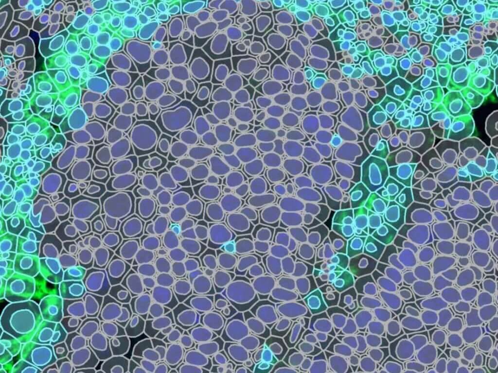
IF Cell Classification


H&E Hepatocellular Ballooning


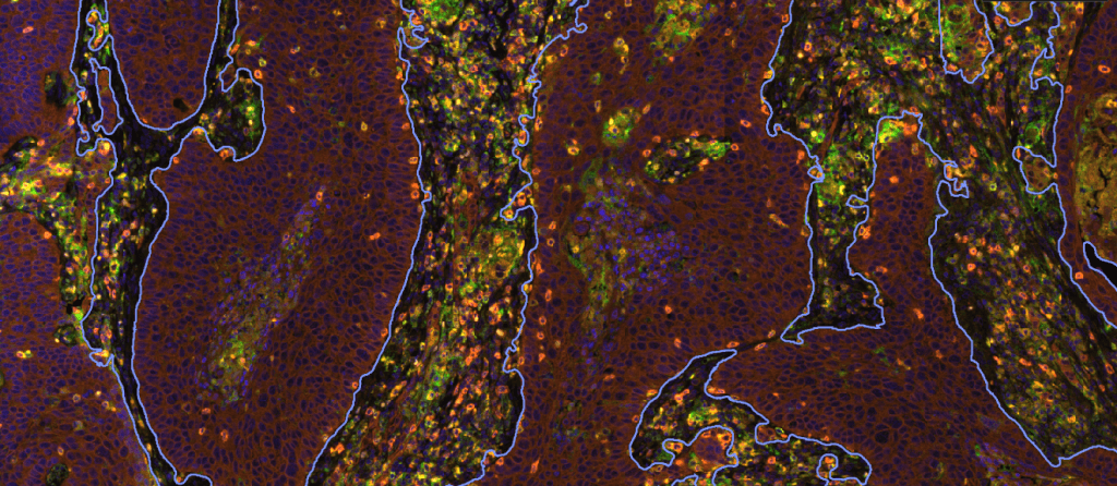
mIF Phenotyping


PR Cell Lines


PDL1 Germinal Center


Invasive Tumor (PDS)


IHC Glomeruli


H&E Lung Metastasis


H&E Intestinal Crypt


Cerebellum LFB


Ki67 Epidermal Hyperplasia
Histopathology Support
Reach out to us early— even during the study design phase— with any questions about tissue sourcing, special staining, H&E, IHC, IF, RNA-ISH, antibody optimization, or other histopathology needs.
We recognize that the foundation of quality data begins even before samples reach our lab. We prioritize collaboration with researchers from the beginning to understand the study scope so we can provide guidance and support as necessary, ensuring that samples are handled with care and precision.
Connect With Us
Your message has been sent
