
Our Histopathology & Image Analysis Solutions
Glint Lab delivers comprehensive histopathology and image analysis services as an extension of your R&D team. From antibody optimization to advanced, AI-driven quantification, every engagement gives you direct access to state-of-the-art technology and expert scientists producing high-quality, actionable data for preclinical and early drug discovery programs.
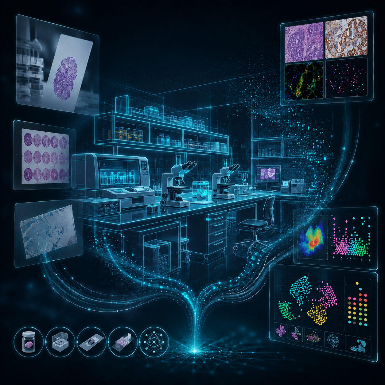
Tailored histopathology and image analysis solutions
Whether you need precision histology, immunohistochemistry, immunofluorescence, or in situ hybridization, our tailored solutions help you extract critical insights from your samples and accelerate your research.
Precision Histology
Routine H&E and special stains built on rigorous tissue processing and embedding.
Learn more →Immunohistochemistry (IHC)
Chromogenic detection of target proteins in FFPE tissue for clear, quantifiable readouts.
Learn more →Immunofluorescence (IF)
Multiplex fluorescent panels for spatial biology and co-expression analysis.
Learn more →In Situ Hybridization (ISH)
RNA and DNA target localization directly within intact tissue morphology.
Learn more →Antibody Optimization and Characterization
Assay development for reliable and reproducible staining and characterizing your novel antibody.
Learn more →Image Analysis
Full-service, AI-driven quantification of cellular and spatial features at scale.
Learn more →Slide Digitization
High-resolution whole-slide imaging at 20x or 40x for brightfield and fluorescent slides.
Learn more →Fractional Computational Services
Extend your team with on-demand access to algorithm development and image analysis expertise.
Learn more →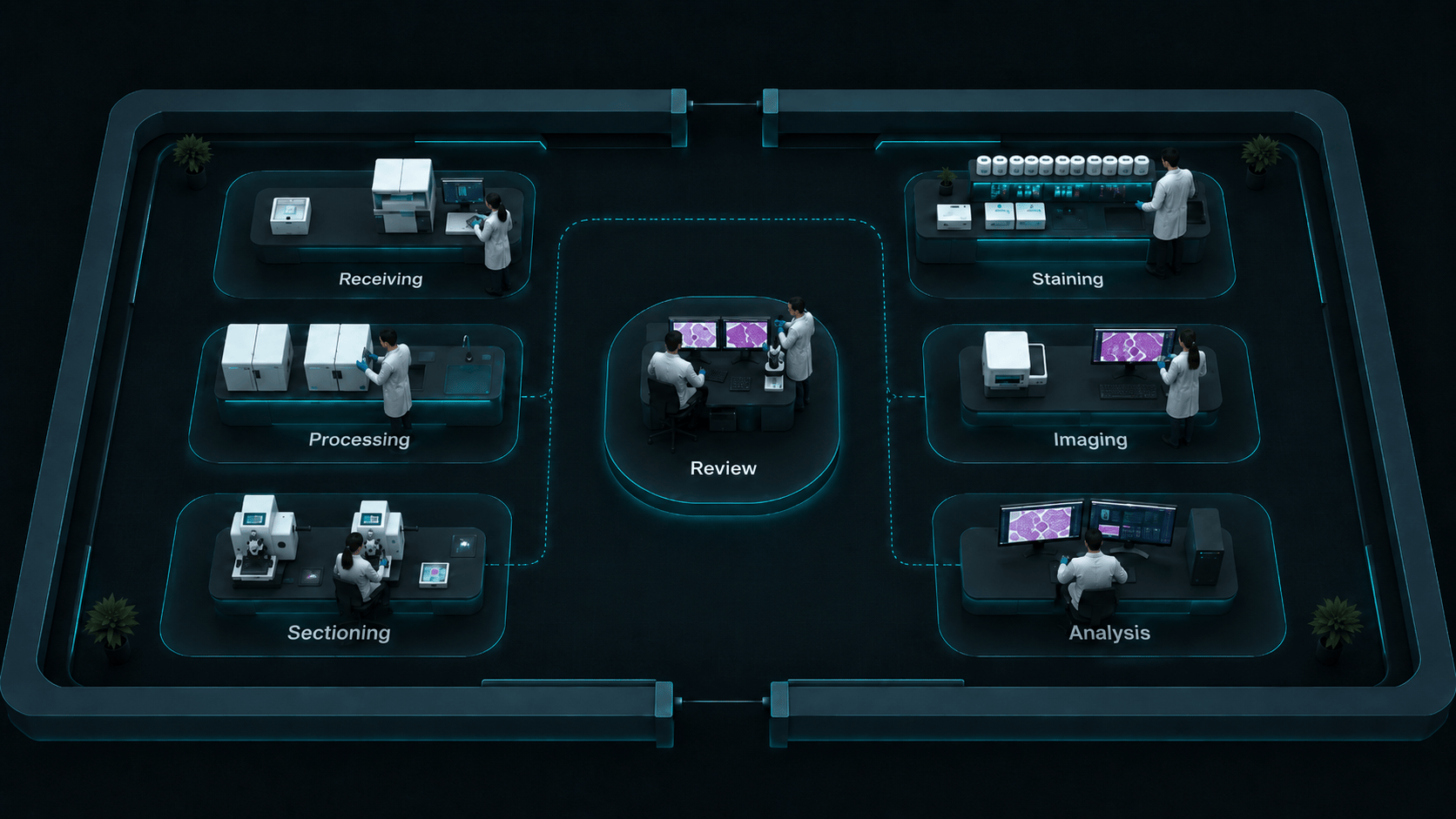
Tissue receiving
Sample intake, accessioning, labeling, and study organization before the tissue enters the workflow.
From tissue to actionable data
Tissue Processing
Formalin-fixed and fresh frozen samples handled to preserve tissue integrity.
Tissue Embedding
Paraffin or optimal cutting temperature (OCT) compound for accurate sectioning.
AI-Assisted Quantification
Advanced algorithms deliver precise, automated analysis via full-service image analysis or fractional computational services.
Expert Evaluation
Comprehensive review by an experienced scientist, a DVM, or a human pathologist.
Comprehensive Reporting
Thorough documentation for iterative scientific discovery, transparency, and accountability.
Built on transparency and collaboration
We prioritize transparency and collaboration to enable better science while making the process easy for you. Our commitment to accuracy and open communication means you can trust us with your critical research needs.
An extension of your R&D
We partner directly with your teams throughout the study, not just at delivery.
Actionable, defensible data
Pathologist-reviewed, quantitative results built for preclinical decision-making.
Flexible engagement
From full-service studies to fractional, on-demand support.
Common questions about our image analysis services
Consult with our histopathology and image analysis experts
See how Glint Lab can support your next preclinical or early drug discovery project.