
Immunofluorescence (IF) Histopathology Services
We offer customized immunofluorescence histopathology services designed to address your specific research requirements, encompassing both single plex and multiplex assays. Depending on your marker or markers of interest, our team will collaborate with you to design optimal staining plan to obtain best results for your tissues of interest, from primary and secondary antibody selections to different multiplex technologies best suited for the markers, species and tissues. We provide high-resolution whole slide images and image analysis to help extract valuable data from your images.


Why Choose GlintLab for IF Histopathology?
- Single-Plex and Multiplex Assays: We design and execute both single-plex IF assays for targeted investigations and multiplex IF assays for complex spatial and co-localization analyses.
- Comprehensive Support: From primary and secondary antibody selection to protocol optimization, we provide end-to-end support for your immunofluorescence studies.
- Multiplex Technology Expertise: Our team is skilled in selecting and implementing the most suitable multiplex technologies for your specific markers, tissue types, and species, ensuring high sensitivity and specificity.
- Advanced Data Outputs: We deliver high-resolution whole-slide images and quantitative image analysis to help you extract actionable insights from your research.
Application Areas of Immunofluorescence:
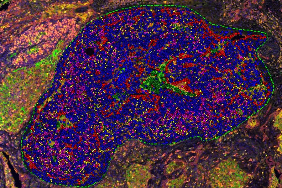
Oncology and Immuno-Oncology
- Immune cell profiling: CD3, CD4, CD8, CD68, PD1, PDL1, FoxP3, and more.
- Tumor microenvironment analysis, including immune cell infiltration and spatial mapping.
Neuroscience
- Detection of key neural markers: GFAP, Iba1, NeuN, S100β, MBP, and others.
- Spatial analysis of neuroinflammatory or degenerative processes.
Dermatology
- Epidermal and dermal marker analysis: COL1A1, Perilipin-1, CD31, and others.
- Automated segmentation of the epidermis and dermis, vessel density and morphology analysis, etc.
Cellular and Molecular Biology
- Tag-based detection: GFP, mCherry, HA Tag, and other fusion proteins.
- Apoptosis markers: Cleaved Caspase 3, TUNEL, and others.
- Analysis of quantitative expression, subcellular localization, and spatial patterns.

Customization for Your Research Goals
Every project at GlintLab begins with a collaborative approach. We work closely with you to:
- Identify the optimal antibodies for your markers of interest.
- Determine the best staining strategies for your tissue types, including FFPE and frozen tissues from human or animal models.
- Optimize protocols to enhance signal detection and minimize background noise for clear, reliable results.

Connect With Us


Related Articles

Brain CNS Neuro Histology CRO Services
The technical considerations discussed in neuro histology cro services align closely with the capabilities described on this pillar page.

Immunofluorescence Image Analysis: Pathology CRO Services
The technical considerations discussed in quantitative immunofluorescence image analysis cro services align closely with the capabilities described on this pillar page.

Quantitative Multiplex image analysis: Pathology CRO Services
The technical considerations discussed in quantitative multiplex image analysis pathology cro services align closely with the capabilities described on this pillar page.

Tissue Fixation Protocol
The technical considerations discussed in tissue fixation align closely with the capabilities described on this pillar page.